Galectin-3 and Frontotemporal Dementia: Emerging Links to Neuroinflammation, Microglial Dysfunction, and Neurodegeneration
What Is Frontotemporal Dementia?
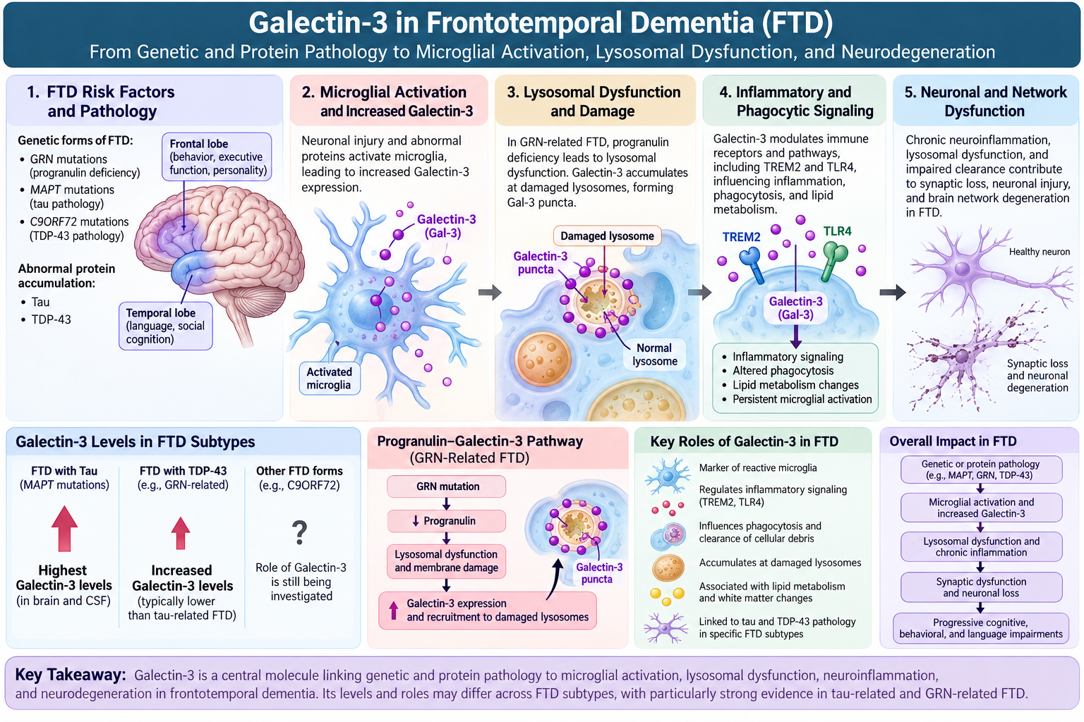
Frontotemporal dementia (FTD) is a group of neurodegenerative disorders characterized by progressive degeneration of the frontal and temporal regions of the brain. These regions are important for behavior, personality, executive function, language, and social cognition. Depending on the brain regions and molecular pathways affected, FTD can present with changes in behavior, language, movement, or combinations of these symptoms.
FTD is biologically diverse. Several different abnormal proteins can accumulate in the brain, with two of the major pathological categories involving:
Tau
TDP-43
Genetic forms of FTD are commonly associated with mutations in genes such as MAPT, GRN, and C9ORF72. In addition to abnormal protein accumulation and neuronal loss, increasing evidence indicates that neuroinflammation and abnormal microglial activity are important components of FTD pathology. One molecule receiving growing attention in this process is Galectin-3 (Gal-3).
What Is Galectin-3?
Galectin-3 is a carbohydrate-binding protein encoded by the LGALS3 gene. Within the central nervous system, Galectin-3 is strongly associated with activated microglia, the resident immune cells of the brain. During neurodegeneration, microglia can change from their normal surveillance state into disease-associated states. Galectin-3 is frequently increased in these reactive microglial populations.
Galectin-3 can influence several biological processes relevant to FTD, including:
microglial activation
inflammatory signaling
phagocytosis
lysosomal function
lipid metabolism
clearance of damaged cellular material
interactions with proteins such as TREM2 and TLR4
responses to neuronal injury
Recent human studies have now demonstrated that Galectin-3 itself is elevated in certain forms of FTD.
Galectin-3 Is Increased in the Brains of Patients With FTD
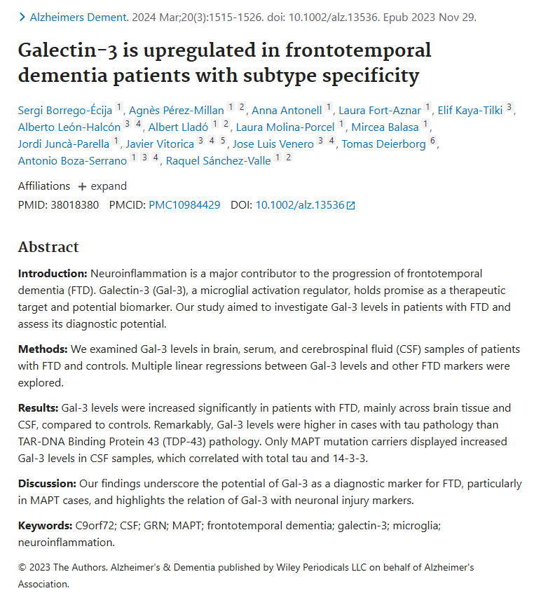
One of the most direct pieces of evidence was published in Alzheimer's & Dementia. Researchers measured Galectin-3 in brain tissue, cerebrospinal fluid (CSF), and serum from patients with FTD and controls. They found that Galectin-3 was significantly increased in FTD, particularly in brain tissue and CSF.
Importantly, the increase was not identical across all types of FTD. Galectin-3 levels were higher in cases with tau pathology than in cases characterized by TDP-43 pathology. Patients carrying MAPT mutations, which cause abnormal tau pathology, also showed increased CSF Galectin-3. CSF Galectin-3 correlated with markers of neuronal injury, including total tau and 14-3-3 proteins. These findings suggest that Galectin-3 may reflect a particular microglial or inflammatory response associated with certain forms of FTD.
Galectin-3 May Differ Between FTD Subtypes as FTD is not a single biological disease. Different genetic and pathological subtypes may activate different neuroinflammatory pathways.
In the human FTD study, Galectin-3 was particularly elevated in FTD associated with tau pathology. The investigators found:
FTD-tau → higher Galectin-3
compared with:
FTD-TDP-43 → relatively lower Galectin-3
Among major genetic forms of FTD, increased CSF Galectin-3 was particularly evident in MAPT mutation carriers. A later systematic review of immune biomarkers in FTD also identified CSF Galectin-3 as one of the biomarkers showing differences among FTD subtypes. This subtype specificity is important because it suggests Galectin-3 may not simply be a general marker of neuronal injury. Instead, it may reflect specific forms of microglial activation occurring in particular molecular forms of FTD.
The Progranulin–Galectin-3 Connection
Another important connection between Galectin-3 and FTD involves progranulin, a protein encoded by the GRN gene. Loss-of-function mutations in GRN reduce progranulin production and are an established genetic cause of FTD. Progranulin plays an important role in:
lysosomal function
lipid metabolism
regulation of inflammation
microglial homeostasis
cellular waste processing
When progranulin is deficient, microglia can develop severe lysosomal and inflammatory abnormalities. Galectin-3 appears to be one of the proteins that becomes strongly elevated during this process.
Galectin-3 Rises Dramatically With Progranulin Deficiency
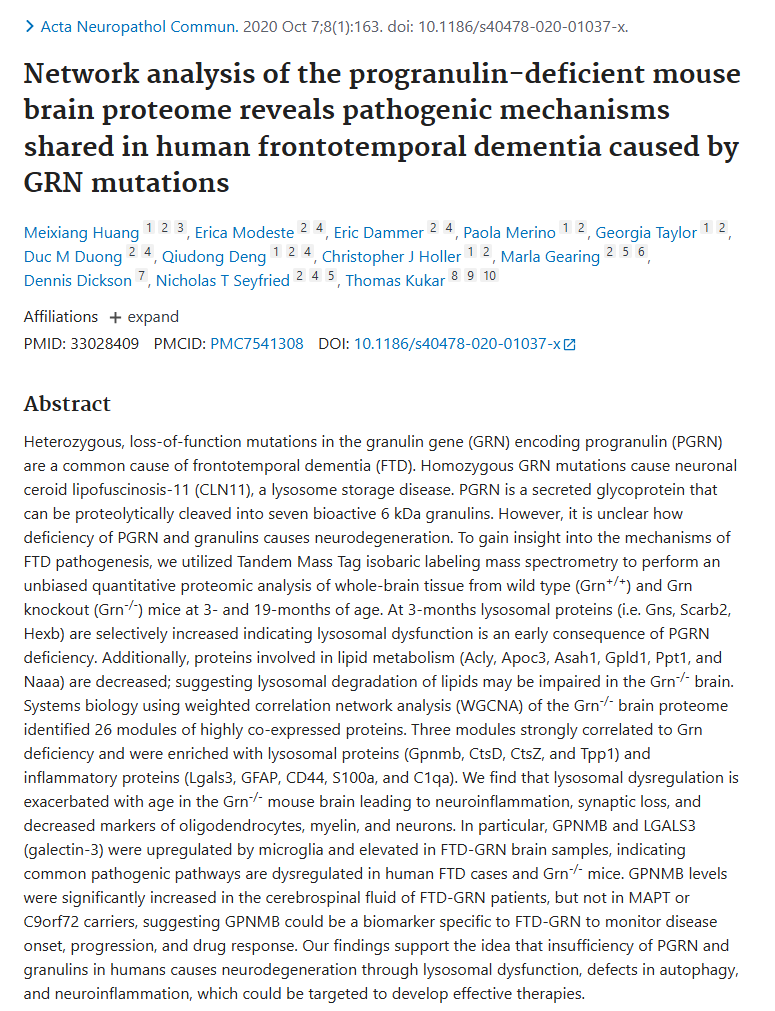
A major proteomic study examined the brains of mice lacking the Grn gene. Researchers found that Galectin-3 was one of the most strongly elevated proteins in the progranulin-deficient brain. In aged Grn-knockout mice, Galectin-3 protein measured by immunoblotting was approximately 21-fold higher than in age-matched control animals. The increase was strongly associated with reactive microglia and became progressively greater with age. Importantly, these findings were not limited to mice.
The investigators also examined human frontal-lobe brain samples from people with FTD caused by GRN mutations. Galectin-3 levels were significantly elevated in FTD-GRN brain tissue compared with cognitively normal controls. This provided one of the first direct links between progranulin deficiency, Galectin-3, microglial activation, and human FTD.
Galectin-3, Microglia, and Neuroinflammation
Microglia normally help maintain the brain by removing cellular debris, damaged proteins, and dysfunctional synapses. In neurodegenerative disease, however, prolonged microglial activation can become maladaptive. Galectin-3 is increasingly recognized as a characteristic protein of certain disease-associated microglial states.
Experimental research suggests that Galectin-3 can interact with important immune receptors such as:
TREM2
TLR4
Through these and other pathways, Galectin-3 can influence inflammatory signaling, phagocytosis, and microglial responses to damaged neurons and abnormal proteins.
Galectin-3 and Lysosomal Dysfunction in FTD
One of the most interesting emerging connections between Galectin-3 and FTD involves the lysosome. Lysosomes are cellular recycling compartments responsible for breaking down:
damaged proteins
lipids
cellular organelles
material engulfed by microglia
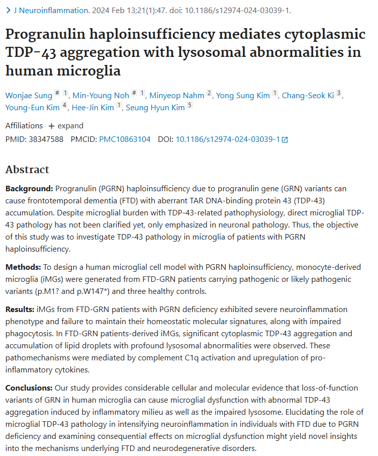
Normal lysosomal function is especially important for microglia because these cells continuously ingest and process cellular debris. GRN-associated FTD is strongly linked to abnormal lysosomal function. A 2024 study generated microglia-like cells directly from patients carrying pathogenic GRN mutations. The researchers found that FTD-GRN microglia had enlarged and dysfunctional lysosomes, abnormal lipid accumulation, impaired phagocytosis, and a strong inflammatory phenotype.
Importantly, LGALS3 — the gene encoding Galectin-3 — was significantly increased in these patient-derived microglia.
Galectin-3 Can Reveal Damaged Lysosomes
Galectin-3 has another unusual property that is particularly relevant to FTD. Normally, Galectin-3 is found in the cytoplasm. When the membrane surrounding a lysosome becomes damaged, carbohydrates normally hidden inside the lysosome become exposed to cytoplasmic Galectin-3. Galectin-3 then accumulates around the damaged lysosome, forming visible Galectin-3 puncta.
For this reason, Galectin-3 is commonly used experimentally as a marker of lysosomal membrane damage. In microglia derived from patients with GRN-associated FTD, researchers observed substantially more Galectin-3 puncta than in control microglia. These Galectin-3-positive structures provided evidence of abnormal lysosomal membrane integrity in the patient-derived cells.
Galectin-3 and TDP-43 Pathology
TDP-43 is another key component of FTD biology. Abnormal accumulation of TDP-43 occurs in a large proportion of FTD cases and is particularly characteristic of GRN-associated FTD. Normally, TDP-43 is primarily located within the cell nucleus. In disease, TDP-43 can move into the cytoplasm, become modified and aggregate, disrupting normal cellular functions.
The 2024 study of patient-derived FTD-GRN microglia found both:
abnormal cytoplasmic TDP-43 accumulation
significant lysosomal abnormalities
in the same disease-associated cells.
These microglia also had significantly increased LGALS3 expression and Galectin-3 puncta, linking Galectin-3-associated lysosomal damage to an environment containing abnormal TDP-43 pathology. The study does not establish that Galectin-3 directly causes TDP-43 aggregation. Instead, it suggests that progranulin deficiency, lysosomal dysfunction, Galectin-3 responses, neuroinflammation, and TDP-43 pathology may be interconnected components of FTD-GRN biology.
Galectin-3, Lipids, and Microglial Dysfunction
Lipid metabolism is another emerging area of interest in FTD. Microglia rely heavily on lysosomes to process lipids from damaged cells and myelin. In progranulin-deficient mice, researchers found abnormalities in lysosomal lipid metabolism accompanied by increasing Galectin-3 expression. Galectin-3-positive microglia were particularly prominent in regions including:
thalamus
cortex
hippocampus
corpus callosum
striatum
The increase in Galectin-3 was also associated with regions showing myelin and white-matter abnormalities.
Because white-matter abnormalities are particularly common in some forms of GRN-associated FTD, this finding raises the possibility that Galectin-3-positive microglia participate in the response to abnormal lipid and myelin processing. Whether Galectin-3 directly drives these changes or is activated in response to them remains under investigation.
Can Correcting Progranulin Deficiency Affect Galectin-3-Related Pathology?
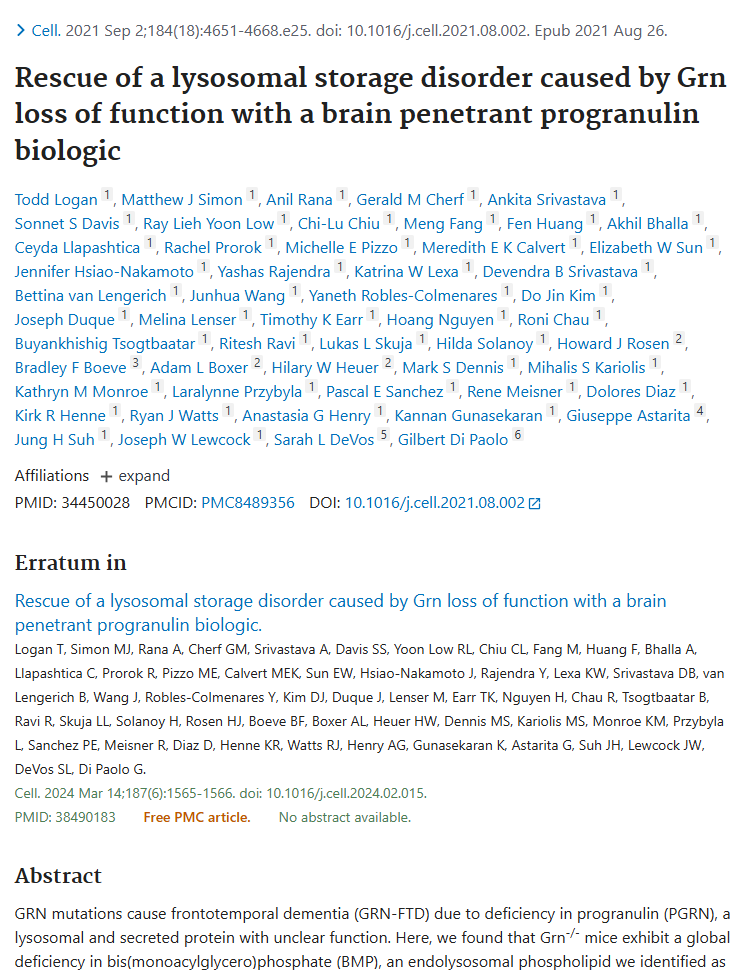
Additional evidence comes from experiments designed to restore progranulin. A study published in Cell tested a brain-penetrant progranulin replacement strategy in progranulin-deficient models. Restoring progranulin corrected several disease-associated abnormalities, including:
lysosomal dysfunction
abnormal lipid metabolism
microgliosis
neuronal damage
The study specifically incorporated Galectin-3 as part of the lysosomal and disease-associated microglial pathology associated with progranulin deficiency.
These findings further connect the progranulin–lysosome–microglia–Galectin-3 axis with FTD-associated disease biology.
Why Is Galectin-3 of Scientific Interest in FTD?
Galectin-3 is particularly interesting because it appears at the intersection of several processes implicated in FTD:
Microglial activation
Galectin-3 is strongly expressed by reactive microglia and is elevated in FTD brain tissue.
Neuroinflammation
Galectin-3 can regulate inflammatory pathways and receptors involved in microglial activation.
Lysosomal dysfunction
Galectin-3 is elevated in progranulin-deficient microglia and accumulates at sites of lysosomal membrane damage.
Abnormal lipid processing
Galectin-3-positive microglial states are associated with changes in lysosomal lipid metabolism and white-matter pathology.
Tau pathology
Human FTD studies have found particularly high Galectin-3 levels in FTD with tau pathology and in MAPT mutation carriers.
TDP-43 pathology
In GRN-associated FTD, elevated Galectin-3 and lysosomal damage occur in microglia displaying abnormal TDP-43 accumulation.
Together, these observations make Galectin-3 an increasingly important molecule for understanding how neuroinflammation and microglial dysfunction may interact with the protein and lysosomal abnormalities underlying FTD.
What Does the Research Show — and What Does It Not Show?
Current research provides direct evidence that Galectin-3 is altered in human FTD.
Studies have demonstrated:
increased Galectin-3 in human FTD brain tissue
increased Galectin-3 in CSF in particular FTD subtypes
especially strong Galectin-3 changes in tau-associated and MAPT-associated FTD
increased Galectin-3 in human FTD-GRN brain tissue
dramatic Galectin-3 upregulation in progranulin-deficient FTD models
increased LGALS3 expression in microglia derived from patients with FTD-GRN
Galectin-3 accumulation around damaged lysosomes in FTD-GRN patient-derived microglia
However, these findings do not yet establish that Galectin-3 is the primary cause of FTD. FTD encompasses several distinct genetic and pathological diseases, and the role of Galectin-3 appears to differ among these subtypes.
It also remains unclear in many settings whether increased Galectin-3 primarily contributes to disease progression, represents a compensatory response to neuronal damage, or performs both functions at different stages of disease. Further research is needed to determine whether modifying Galectin-3 activity can alter the course of FTD in humans.
Scientific Foundations: The Evidence for Galectin-3 and FTD
-
![]()
Journal: Alzheumers Dement.
Galectin-3 is upregulated in frontotemporal dementia patients with subtype specificity (2024)
-
![]()
Journal: Acta Neuropathol Commun.
Network analysis of the progranulin-deficient mouse brain proteome reveals pathogenic mechanisms shared in human frontotemporal dementia caused by GRN mutations (2020)
-
![]()
Journal: Cell
Rescue of a lysosomal storage disorder caused by Grn loss of function with a brain penetrant progranulin biologic (2021)
-
![]()
Journal: Neuroinflammation
Progranulin haploinsufficiency mediates cytoplasmic TDP-43 aggregation with lysosomal abnormalities in human microglia (2024)