The Cellular Anchor: How Galectin-3 Roots, Fuels, and Inflames Endometriotic Lesions
Introduction
The Shift in Research For decades, endometriosis has been viewed almost exclusively as a estrogen-dependent hormonal disorder characterized by retrograde menstruation. However, surgical removal of lesions and hormonal suppression therapies often fail to prevent recurrence or completely alleviate debilitating pelvic pain. Leading research shows that endometriosis is fundamentally an inflammatory and tissue-remodeling disease. At the crossroad of ectopic cell survival, blood vessel formation (angiogenesis), and scarring is a critical carbohydrate-binding protein: Galectin-3 (Gal-3).
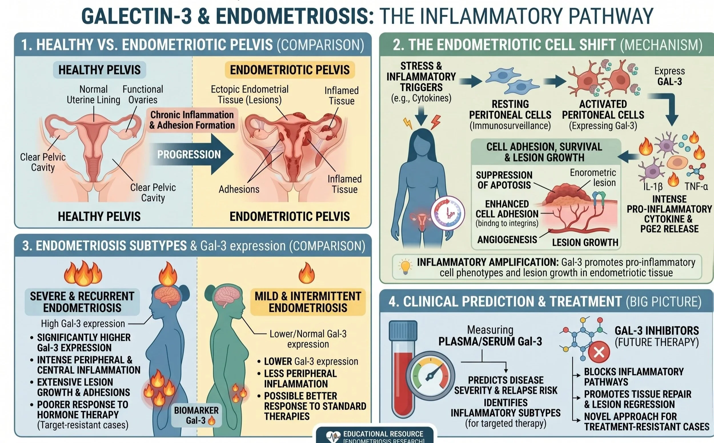
What is Gal3 in Endometriosis? Galectin-3 (Gal-3) is a pro-inflammatory lectin secreted by peritoneal macrophages, vascular endothelial cells, and ectopic endometrial cells. While Gal-3 normally regulates tissue repair and immune response, its pathological overexpression in the pelvic cavity creates a permissive environment that allows endometrial tissue to attach outside the uterus, evade immune clearance, construct new blood vessel networks, and form painful fibrotic adhesions.
How Gal3 Drives Endometriotic Lesion Progression:
1. Cell Adhesion & Tissue Invasion (Anchoring the Lesion)
Ectopic endometrial cells that enter the pelvic cavity must adhere to the peritoneal lining to survive.
The Gal3 Effect: Extracellular Gal-3 binds to $\beta$-galactoside sugars on surface integrins and extracellular matrix (ECM) proteins (such as fibronectin and laminin). This acts as a molecular "glue," facilitating the attachment of shedding endometrial cells to the peritoneum and aiding their invasion into pelvic structures.
2. Angiogenesis: Building the Blood Supply
For ectopic lesions to thrive and grow, they require a dedicated vascular network to deliver oxygen and nutrients.
The Gal3 Effect: Gal-3 directly stimulates endothelial cell migration and capillary tube formation. It interacts synergistically with Vascular Endothelial Growth Factor (VEGF), accelerating neovascularization and turning small surface implants into deeply vascularized, active endometriotic lesions.
3. Immune Evasion & Inflammatory Amplification
Normally, scavenger macrophages in the peritoneal fluid clear refluxed endometrial tissue.
The Gal3 Effect: Ectopic cells secrete Gal-3 to modulate local immune surveillance, suppressing apoptosis (programmed cell death) of the lesion cells. Simultaneously, Gal-3 triggers peritoneal macrophages to release inflammatory mediators—including Interleukin-1 beta (IL-1beta), Tumor Necrosis Factor-alpha (TNF-alpha), and Prostaglandin E2 (PGE2)—inducing local hyperalgesia and pelvic nerve sensitization.
4. TGF-b Signaling & Fibrotic Adhesions
The chronic cycle of bleeding and inflammation leads to irreversible scar tissue (adhesions) that binds pelvic organs together.
The Gal3 Effect: Gal-3 directly activates TGF-$\beta$/Smad signaling pathways in peritoneal fibroblasts. This drives myofibroblast differentiation and excessive collagen deposition, resulting in severe pelvic adhesions, organ tethering, and deep infiltrating endometriosis (DIE).
Therapeutic Potential: Target Gal3 to Halt Endometriosis
Standard hormonal therapies (such as GnRH agonists or progestins) put patients into temporary menopausal states and carry significant side effects, yet lesions often recur post-treatment.
Blocking Galectin-3 presents a novel, hormone-independent therapeutic avenue:
Lesion Regression: Inhibiting Gal-3 disrupts integrin-mediated adhesion, preventing ectopic cells from attaching to the peritoneal surface.
Anti-Angiogenic Effect: Gal-3 inhibition starves established lesions by blocking VEGF-driven blood vessel formation.
Pain & Fibrosis Reduction: Suppressing Gal-3 halts the chronic release of inflammatory prostaglandins and attenuates TGF-beta-mediated pelvic scar formation.
Scientific Foundations: The Evidence for Galectin-3 and Endometriosis
-
![]()
Journal: Mol Cell Endocrinol
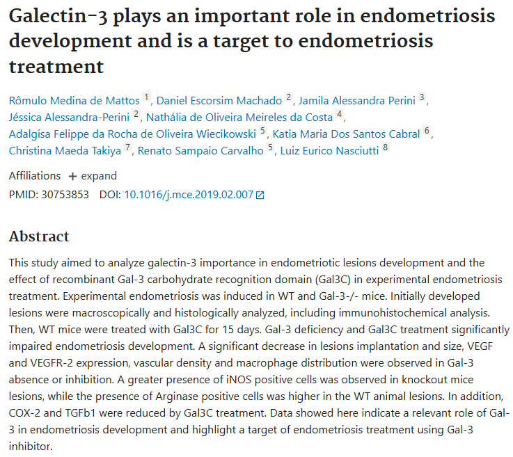
Galectin-3 plays an important role in endometriosis development and is a target to endometriosis treatment (2019)
-
![]()
Journal: Gynecol Endocrinol
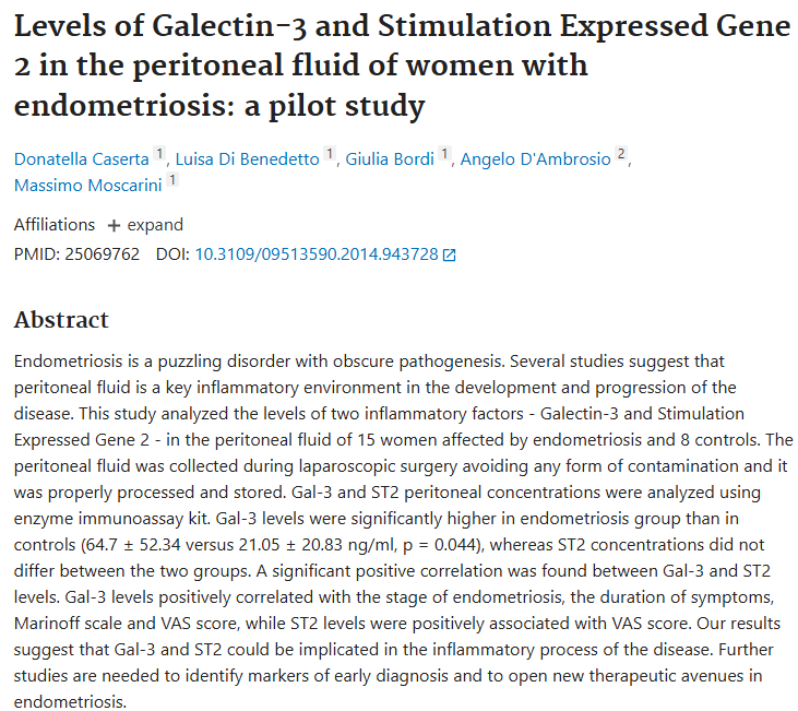
Levels of Galectin-3 and Stimulation Expressed Gene 2 in the peritoneal fluid of women with endometriosis: a pilot study (2014)
-
![]()
Journal: Int J Molecular Sciences
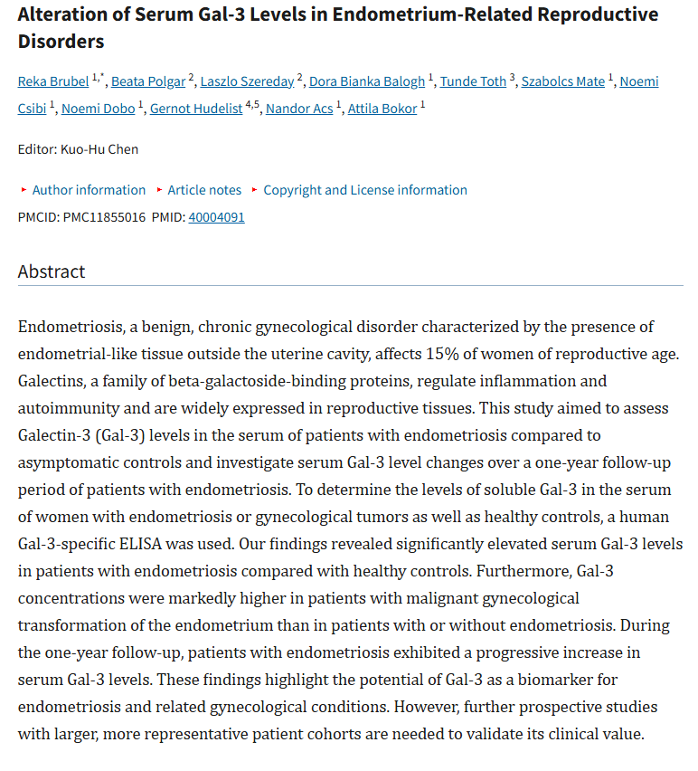
Alteration of Serum Gal-3 Levels in Endometrium-Related Reproductive Disorders (2025)
-
![]()
Journal: Peer J
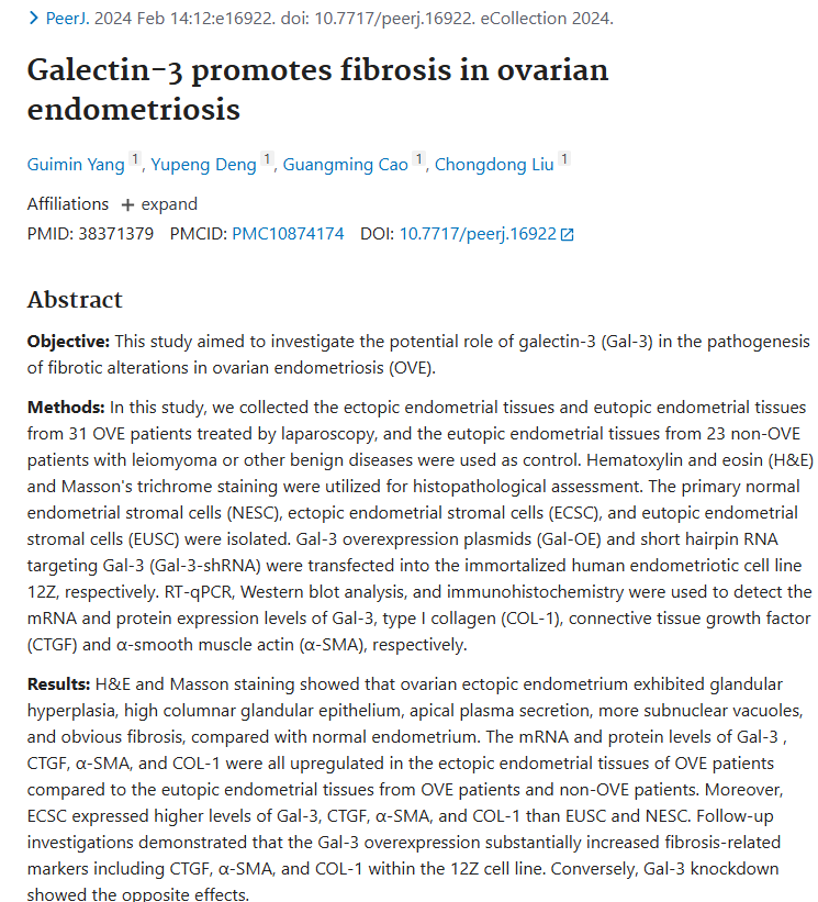
Galectin-3 promotes fibrosis in ovarian endometriosis (2024)