The Early Alarm: Is Galectin-3 the Amplifier Driving Encephalitis?
Introduction
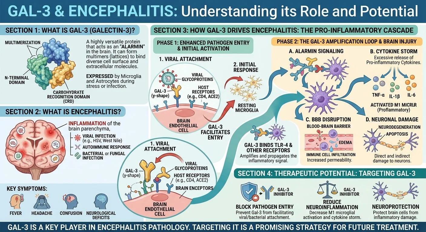
What it is: Encephalitis is inflammation of the brain parenchyma, a serious and potentially life-threatening condition that can cause seizures, confusion, personality changes, and coma.
Two main types: Infectious and autoimmune are the two main divisions.
Infectious encephalitis: Caused by viruses invading the brain, such as herpes simplex virus (HSV), enteroviruses, West Nile virus, and Japanese encephalitis virus. Mosquitoes and ticks can transmit these viruses. Symptoms develop quickly, over days to weeks.
Autoimmune encephalitis: The immune system mistakenly attacks the brain, often targeting neuronal surface proteins (e.g. NMDA receptor, LGI1). It can be triggered by infection or tumor, but in about 90% of cases the cause is not found. Symptoms can develop over weeks to months and include personality change, memory loss, psychosis, seizures, and unusual movements.
Early diagnosis and treatment are critical — treatment is urgent and usually in hospital with antivirals, steroids, or immunotherapy depending on type.
How Galectin-3 Is Involved — Mechanism of Action
The core idea: Galectin-3 (Gal-3) is absent in the healthy brain but appears during ongoing brain inflammation. It is secreted by microglial cells and binds to the TLR4 receptor, amplifying the inflammatory reaction in a self-sustaining loop.
Here’s how Gal-3 drives encephalitis pathology:
1. Early alarm in viral encephalitis: After acute viral inoculation (EMCV mouse model), Gal-3 is up-regulated in degenerated lesions in cerebellum, hippocampus, thalamus and cerebral hemisphere by 96h. Iba-1 positive microglia are morphologically activated within and around the focus. Strikingly, microlesions with Gal-3-positive cells are detectable as early as 48h — suggesting Gal-3 is a key early mediator between viral infection and neuronal degeneration.
2. TLR4 amplification loop: Activated microglia release Gal-3, which binds TLR4 on microglia, driving NF-κB and JAK-STAT signaling and production of TNF-α, IL-1β, IL-6, and IFN-γ. More Gal-3 is produced, further activating microglia — a feed-forward cytokine storm.
3. Phagocytosis of neurons and BBB disruption: Gal-3 binds desialylated neurons, opsonizing them for microglial phagocytosis, and may increase blood-brain barrier permeability and recruit peripheral monocytes via chemoattractant properties.
4. Autoimmune angle (EAE model): In experimental autoimmune encephalomyelitis, Gal-3 deficiency results in less severe disease with reduced monocyte infiltration, IL-17 and IFN-γ in the CNS. This supports a pro-inflammatory role relevant to autoimmune encephalitis.
Therapeutic hypothesis: If Gal-3 is the amplifier, blocking it (with Gal-3 inhibitors / antibodies) could calm microglia, reduce cytokine release, and limit neuronal loss — similar logic to your depression page, but here applied to acute infectious and autoimmune brain inflammation. Most data so far are preclinical (mouse models).
Scientific Foundations: The Evidence for Galectin-3 and Encephalitis
-
![]()
Journal: Affective Disorders
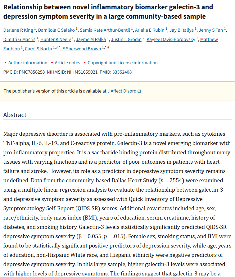
Relationship between novel inflammatory biomarker galectin-3 and depression symptom severity in a large community-based sample (2022)
-
![]()
Journal: International Journal Molecular Sciences
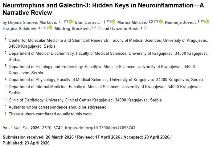
Neurotrophins and Galectin-3: Hidden Keys in Neuroinflammation—A Narrative Review (2026)
-
![]()
Journal: Human Psychopharmacology-Clinical and Experimental
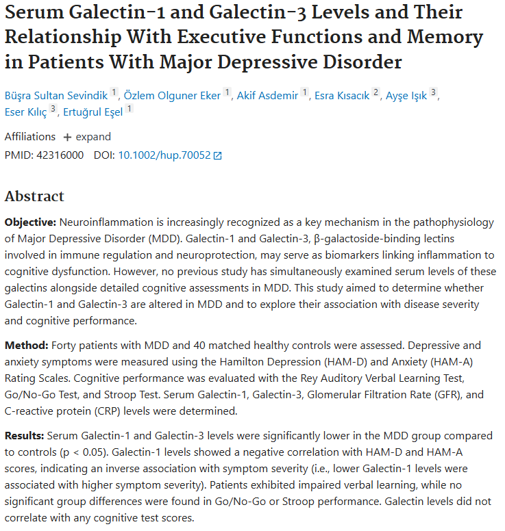
Serum Galectin-1 and Galectin-3 Levels and Their Relationship With Executive Functions and Memory in Patients With Major Depressive Disorder (2026)